Diana Arseni from the MRC Laboratory of Molecular Biology, UK
Diana graduated with an MSci in Biomedicine from the University of Lancaster in 2016. During her PhD at the University of Glasgow and Astrazeneca, she investigated mechanisms through which microglia mediate tissue damage in the context of multiple sclerosis. In 2020, she started her postdoc at the MRC LMB, in the lab of Ben Ryskeldi-Falcon, where her work focused on elucidating the structure of TDP-43 filaments from neurodegenerative diseases using cryo-electron microscopy. In November 2024, she started her own research group in the Neurobiology Division at the MRC LMB. Her group studies mechanisms of brain health, ageing and disease focusing on the lysosomal protein transmembrane protein 106B (TMEM106B) which forms amyloids in the brain in an age-dependent manner.
Talk Abstract
The ABC of TDP-43
Abnormal assemblies of TDP-43 in neurons and glia are the pathological hallmark of amyotrophic lateral sclerosis (ALS) and multiple types of frontotemporal lobar degeneration (FTLD). Mutations in the TDP-43 gene, TARDBP, can cause ALS and FTLD,and the temporospatial accumulation of TDP-43 assemblies correlates with neurodegeneration, indicating a causative role for TDP-43 assembly in disease. TDP-43 assemblies are also common co-pathologies in other diseases, including Alzheimer’s, Parkinson’s and Huntington’s. The structural and molecular mechanisms of TDP-43 assembly in disease are poorly understood. We developed a protocol to isolate assembled TDP-43 from the brains of patients with ALS and FTLD and determined their structures using cryo-electron microscopy (cryo-EM). We found that TDP-43 assembles into amyloid filaments in these diseases. The ordered filament cores are comprised of the first half of the TDP-43 low-complexity domain and adopt distinct filament folds in different neurodegenerative conditions. These brain-derived filament folds show no similarity to TDP-43 filament folds formed in vitro. The structures, in combination with mass spectrometry, led to the identification of two new post-translational modifications of assembled TDP-43, citrullination and mono-methylation of R293, and suggest that they may facilitate filament formation and observed structural variation within individual filaments. Unexpectedly, the structures also revealed that in specific cases TDP-43 can also co-assemble with Annexin A11 in heteromeric amyloid filaments. The structures of TDP-43 amyloid filaments from ALS and FTLD guide mechanistic studies of TDP-43 assembly, as well as the development of diagnostic and therapeutic compounds for TDP-43 proteinopathies.
Morgan Beeby from the Imperial College London, UK
Morgan studied for a BSc at the University of Birmingham. A summer project refining a crystal structure inspired a move to a PhD under Todd O. Yeates at UCLA where he trained in crystallography and bioinformatics to understand how hyperthermophiles have adapted to survive at high temperatures, and the genomic basis for self-assembly of bacterial microcompartments and analogous structures. To better understand principles of biological self-assembly and evolution, he next trained in electron cryo-tomography with Grant J. Jensen at Caltech, working on projects including cataloging the diversity of bacterial flagellar motors, nanoscale motors that spin helical filaments to act as propellors. He started his own lab in 2013 using electron cryo-tomography as the core technique to continue work that uses the structural diversity of flagellar motors to shed light upon basic principles of molecular assembly, function, and evolution.
Talk Abstract
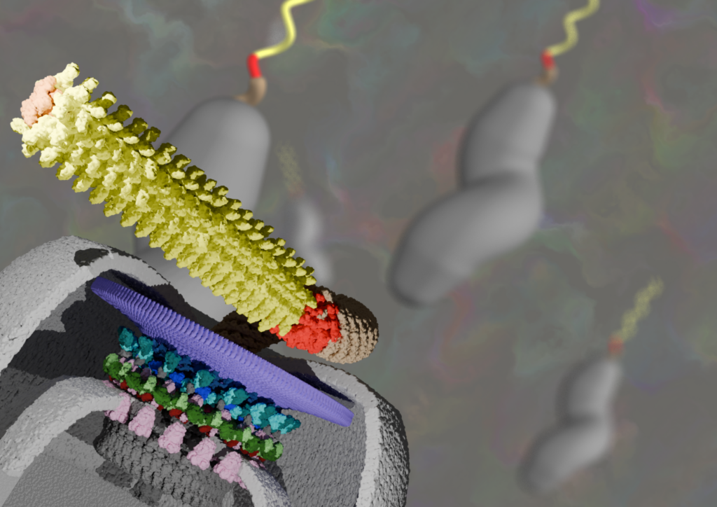
Electron cryo-microscopy of bacterial flagellar motors in situ reveals clues to their evolution
Bacterial flagellar motors are excellent case studies for understanding the mechanisms of molecular machines, and their evolution. Progress has been hampered by relatively low resolutions that fail to provide sufficient constraints for unambiguous molecular interpretation. Here I describe our recent work using electron cryo-microscopy to attain subnanometre resolutions of flagellar motors in situ, and the biological insights that this enables.
Nicholas Dale from the University of Warwick, UK
Following a degree in Zoology from Cambridge, Nicholas obtained a PhD in Neuroscience at the University of Bristol with Prof Alan Roberts. He was a post-doctoral fellow with Prof Sten Grillner at the Karolinska Institute, Stockholm, and then with Dr Eric Kandel at Columbia University, New York. He returned to the UK in 1989 to hold a Royal Society University Research Fellowship at Bristol. Nicholas then gained a Royal Society Locke Research Fellowship and held this initially at Bristol but moved to the University of St Andrews in 1995. In 2000, he moved to his current position as the Ted Pridgeon Professor of Neuroscience at the University of Warwick. In 2015, he gained a Royal Society Wolfson Research Merit Award for his work on CO2 chemosensing.
Talk Abstract
CO2–sensing via connexins: structural biology, physiology and evolution
Connexins canonically form gap junction channels that allow the passage of ions and small molecules between adjacent cells. However, they can also act as unopposed hemichannels, opening to the extracellular medium. Hemichannels of the three most widely expressed connexins in the human body are directly gated by CO2. Although CO2 is often regarded as a metabolic waste product, the ubiquity of CO2-sensitive connexin hemichannels suggests that CO2 may also be an important physiological signal. We have discovered that CO2 gates connexin hemichannels via a non-enzymatic carbamylation mechanism at an ancient evolutionarily conserved motif that is present in all CO2-sensitive connexins. I shall outline our understanding of how CO2 opens connexin channels and discuss some of the physiological/behavioural functions of connexin-mediated CO2 signalling. I will put forward our evidence for CO2acting as a specific cell-to-cell signal akin to a neurotransmitter. While the connexin gene is considered to be a chordate innovation, I will present our evidence that connexins have deep evolutionary origins from connexin-like domains in genes relating to innate immunity in Cnidaria and Protostomes.
Giovanni D’Angelo from EPFL, Switzerland
Giovanni D’Angelo graduated in 2003 with a MSc in Medical Biotechnology from the University of Naples, Italy and obtained his PhD in Cell Biology in 2008 from the Consorzio ‘Mario Negri’ SUD, Santa Maria Imbaro, Italy. For his postdoctoral training, he moved to the Telethon Institute for Genetics and Medicine in Naples, Italy to study sphingolipid metabolism and intracellular lipid trafficking. In 2012, Giovanni moved to the Institute of Protein Biochemistry, at the National Research Council of Italy in Naples as a principal investigator. In 2018 Giovanni moved to the Swiss Federal Institute of Technology in Lausanne (EPFL) where he is now Associate Professor and Kristian Gerhard Jebsen Chair on Metabolism, Giovanni’s main interest is understanding the meaning of compositional variability in cell membranes by studying the mechanisms by which the lipid composition is determined.
Talk Abstract
The lipidomic architecture of the mouse brain
Lipids are fundamental components of the brain, crucial for synaptic transmission and signal propagation. Altered brain lipid composition is associated with common and rare neuropathologies, yet, the spatial organization of the mammalian brain lipidome remains insufficiently characterized compared to other modalities. Here, we mapped the membrane lipid architecture of the adult mouse brain at micrometric scale, across sexes, and during pregnancy. This Lipid Brain Atlas reveals that lipids define a fine-grained biochemical structure that aligns with functional anatomy. Membrane lipid spatial heterogeneity clusters into territories, which we termed lipizones. Lipizones partially mirror cell type territories, but also capture distal axon terminals. Through lipizones, (i) we reveal the organizing principles of the gray matter lipidome, related to connectivity and cytoarchitecture; (ii) we discover a new axis of oligodendrocyte heterogeneity in the white matter; (iii) and we find biochemical zonation in the choroid plexus and in the ventricular walls. We show that this lipidomic architecture can adapt to changing physiological needs. In the brain of pregnant females, the white matter is metabolically activated and the outer cortex is reorganized. These results are a foundational resource (https://lbae-v2.epfl.ch/), poised to reshape our understanding of lipids in brain development, physiology, and pathology.

Shannan Foylan from the University of Strathclyde, UK
Shannan is a postdoctoral research scientist at the Strathclyde Institute for Pharmacy and Biomedical Sciences, Glasgow UK. She has multidisciplinary research interests in molecular biology, transporter biology in disease and the optical design and implementation of state-of-the-art high and super-resolution optical imaging. Shannon completed her PhD in 2023 in the Department of Physics at the University of Strathclyde, developing several novel high throughput imaging methods with improved axial resolution for quicker imaging of larger cell populations. Her current research focuses on elucidating the mechanism for dispersal of the glucose transporter molecule GLUT4 in the plasma membrane of insulin responsive cells, with the long-term aim of understanding the impact of insulin-resistance/diabetes on this novel aspect of GLUT4 cell biology.
Talk Abstract
After a meal, rising blood glucose triggers insulin release from the pancreas, promoting glucose uptake into tissues via insulin-responsive molecular glucose transporters (GLUTs). In muscle and adipose cells, GLUT4 is the primary transporter responsible for this process, and defects in its regulation are hallmarks of insulin resistance and Type-2 diabetes. While intracellular trafficking of GLUT4 is well characterised, less is known about its organisation and diffusion within the plasma membrane (PM). Previous work has shown that GLUT4 forms stationary clusters under basal conditions but redistributes into diffusing monomers following insulin stimulation, a process requiring the scaffold protein EFR3.
To investigate GLUT4 nanoscale organisation, we applied both Single Molecule Localisation Microscopy and Ultrastructure Expansion Microscopy (U-ExM) to adipocytes and cardiomyocytes. We have established a pipeline for applying U-ExM to murine-derived adipocyte cell monolayers, achieving four-fold isotropic expansion and enabling large-volume, super-resolution imaging of GLUT4 using standard diffraction-limited systems. This approach provides an accessible alternative to costly super-resolution platforms for metabolic studies.
Complementary live-cell imaging and Fluorescence Correlation Spectroscopy revealed distinct dynamic behaviours of clustered versus dispersed GLUT4 molecules. Together, our findings uncover differences between basal and insulin-stimulated states and provide tools to study GLUT4 dynamics and insulin responsiveness in situ in metabolic tissues.

Stephan Gruber from the University of Lausanne, Switzerland
Stephan is an Associate Professor in the Department of Fundamental Microbiology at the University of Lausanne. His research integrates structural biology, biochemistry, and molecular genetics to elucidate how SMC DNA-folding machines drive key cellular processes in genome maintenance and immunity. Stephan earned his PhD in 2005 from the University of Vienna, where he studied cohesin and chromosome segregation in yeast under the supervision of Kim Nasmyth. He subsequently transitioned to bacterial systems, expanding his expertise in chromosome biology as a postdoctoral fellow with Jeff Errington at Newcastle University. After establishing an independent research group at the Max Planck Institute of Biochemistry in Munich in 2010, he relocated his laboratory to the University of Lausanne in 2016.
Talk Abstract
From Folding to Fending: How a chromosome-organizing machine combats foreign DNA
Structural Maintenance of Chromosomes (SMC) complexes are nearly ubiquitous molecular motors that organize genomes through ATP-driven, stepwise folding of DNA, a process known as DNA loop extrusion. While they are best characterized for their roles in chromosome organization and segregation, recent work has revealed that SMC complexes can also be co-opted for immunity. Among these, the Wadjet (JetABCD) complex functions as an elegant anti-plasmid defence system in bacteria and archaea. We show how Wadjet exploits the mechanics of loop extrusion to distinguish self from non-self DNA without relying on sequence-specific recognition or DNA modification. The complex comprises an SMC sensor module (JetABC) and an endonucleaseeffector (JetD). It binds DNA non-specifically and reels it in symmetrically. On host chromosomes, extrusion proceeds effectively indefinitely due to the large size of the DNA molecule. In contrast, when Wadjet encounters smaller, circular DNA—such as a plasmid or a prophage genome—it extrudes the entire length of the DNA until it stalls. This “total extrusion” functions as a DNA size-sensing mechanism, triggering a conformational change that activates the JetD nuclease to cleave and neutralize the foreign DNA. By coupling a chromosome-organizing motor with a nuclease effector, Wadjet illustrates a remarkable evolutionary transition: the repurposing of a fundamental genome-folding machine into a highly specific defence weapon.

Sir Steve Jackson from the Cancer Research UK Cambridge Institute, UK
Sir Stephen (Steve) Jackson FRS, FMedsci, FAACR is a Professor of Biology at the University of Cambridge and Senior Group Leader at the CRUK Cambridge Institute (CI). After graduating in biochemistry from the University of Leeds, Steve carried out his PhD at Imperial College London and the University of Edinburgh, then carried out post-doctoral training at UC Berkeley. He returned to the UK in 1991 to set up his research group in what is now the Gurdon Institute, and moved to the CI in 2022. He has received various national and international prizes, and was awarded a knighthood for his services to innovation and research in 2023.
Talk Abstract
Cellular responses to DNA damage: mechanistic insights and medical implications
Steve’s academic research has identified key principles by which cells respond to and repair DNA damage and defined how their dysfunction yields cancer and other age-related diseases. In 1997, Steve founded the drug-discovery company KuDOS Pharmaceuticals, which developed and took into first patients the PARP inhibitor drug olaparib (LynparzaTM), now marketed worldwide for certain ovarian, breast, pancreatic and prostate cancers. In 2010, Steve founded Mission Therapeutics to exploit advances in protein ubiquitylation and deubiquitylation to derive new medicines. In 2018, he conceived of and co-founded Adrestia Therapeutics, which in 2023 was acquired by Insmed Inc.
His laboratory in the CI focuses on defining mechanisms by which cells detect, signal the presence of and repair DNA damage, and exploring how this knowledge might lead to new therapeutic approaches. This work involves biochemistry and precision cell biology underpinned by CRISPR-based genome engineering and genetic screens.

Alex Johnson from the University of Exeter, UK
During his PhD (with Prof Mike Cousin, University of Edinburgh) investigating synaptic vesicle recycling by post-translational modification, Alex became interested in how other model systems can complete the same fundamental cellular processes with evolutionarily distinct protein machinery and overcoming distinct biomechanical properties. Following this interest, he undertook post doc positions (with Dr. Gregory Vert, CNRS and Prof Jiri Friml, IST Austria) to investigate the molecular mechanisms of plant clathrin-mediated endocytosis, finding it operated differently from predictions based upon mammal and yeast models, and in a surprisingly mechanistically actin-independent nature. This highlighted the significance of biomechanics, so he did a post doc in physics (with Dr. Kareem Elsayed, Medical University of Vienna) to optimise contact- and label-free optical tools to probe biomechanics in living samples. Now at Exeter, Alex is combining cell biology, physics, plants and diatoms to uncover how clathrin-mediated trafficking underlies cell-environmental interactions in photosynthetic model systems.
Talk Abstract
From land and sea: Clathrin-mediated cellular sensitivity in photosynthetic organisms
Climate change is negatively impacting the ability of plants and marine algae to grow, affecting life on Earth as together they provide our global staple food, produce ~70% of our oxygen and underpin terrestrial and marine ecosystems. Therefore, it is critical to understand the cell biology underpinning how cells perceive their environments to identify novel climate-resilience targets. Clathrin is a fundamental eukaryotic protein, and through clathrin-mediated endocytosis, it regulates the number of receptors on the cell surface and controls how extracellular materials (including pathogens, toxins, and agrichemicals) enter the cell, thus directly regulating organism environmental sensitivity. However, almost all our understanding of its molecular details has been derived from mammalian and yeast cells. My work has demonstrated that plant CME is evolutionarily and mechanistically distinct; overcoming high turgor independently of actin and relying on proteins absent from mammalian and yeast genomes. Together, this highlights the need to refine our principles of CME across the tree of life. I will present our work updating working models of plant CME, including the development of quantitative imaging tools to probe CME at multiple scales in intact plants. I will then present how we are transferring these approaches to diatoms, an evolutionarily distinct photosynthetic model system, to compare CME across photosynthetic lineages and identify unique features that could be targeted to tune environmental responses.

Joergen Kornfeld from the MRC Laboratory of Molecular Biology, UK
Dr. Joergen Kornfeld is a connectomics researcher and neuroscientist at the MRC Laboratory of Molecular Biology in Cambridge. His group builds a vertebrate brain scale volume electron microscopy pipeline and develops machine learning methods to automate connectome extraction and analysis, studying how the zebra finch stores its learned song in synaptic wiring. He studied molecular biology as an undergraduate at the University of Heidelberg and later computational biology at ETH Zurich. For his PhD, he trained with Winfried Denk at the Max Planck Institute in Heidelberg, followed by a postdoc with Michale Fee at MIT before starting his lab at Max Planck in Munich. In 2024, he moved to the LMB to establish the Connectomics of Learned Behavior group.
Talk Abstract
How Connectomics Is Slowly Revolutionising Neuroscience
Sydney Brenner founded the field of synaptic-resolution connectomics in Cambridge with the C. elegans connectome and showed that a complete circuit map fundamentally changes the questions we ask about brains. Today, whole-brain fly connectomes are repeating that history, with many species to come in the next few years. I will describe how we are scaling to vertebrate brains, focusing on the zebra finch as a model for complex learned behaviour. The talk covers two practical bottlenecks: high-throughput image acquisition and automated reconstruction, in which multibeam SEM and modern deep networks make previously impossible samples tractable while leaving open challenges in quality control and in making petabyte-scale data truly queryable. With examples from the songbird, I will sketch how connectomes let us test circuit hypotheses and outline our long-term goal: to perform a post-mortem read-out of a complex memory from synaptic wiring and thereby test our understanding of vertebrate brain function bottom up.
Fides Zenk from EPFL, Switzerland
Fides Zenk is an Assistant Professor at the Brain Mind Institute of EPFL, where she holds the NeuroNA Chair of Epigenomics and Neurodevelopmental Disorders. Her research investigates how epigenetic regulation shapes human brain development and how its disruption leads to neurodevelopmental disease. During her PhD at the Max Planck Institute, she uncovered fundamental principles of chromatin regulation, including mechanisms of histone modification inheritance and their impact on genome organization. In her postdoctoral work, she developed single-cell epigenomic approaches to map the regulatory landscape of developing tissues and reconstruct developmental trajectories. In her current work, her lab combines human stem cell–derived brain organoids with single-cell genomics to define how epigenetic programs are established during human neurodevelopment and how their failure contributes to disease.
Talk Abstract
Reconstructing Human Neurodevelopment with Single-Cell Epigenomics
How do genetically identical cells give rise to the extraordinary diversity of cell types in the human brain? A tightly controlled series of fate restrictions from pluripotent progenitors drives this process, guided by epigenetic mechanisms that regulate gene activity and associated regulatory elements. However, studying these mechanisms during early human brain development has remained challenging. In our lab, we develop single-cell technologies to map epigenetic modifications and resolve cell fate decisions at high resolution. Using these approaches, we profiled histone modifications (H3K27ac, H3K27me3, and H3K4me3) in human central nervous system organoids across a developmental time course and reconstructed the epigenomic trajectories underlying cell identity acquisition from human pluripotency. We analyzed transitions from pluripotency to neuronal and glial terminal states, as well as differentiation from progenitors to retinal and brain regional identities through the neuroepithelium. We found that cell fate decisions could be predicted by dynamic switching between repressive and activating epigenetic modifications. Furthermore, we established a temporal census of regulatory elements and transcription factors and positioned them within gene regulatory networks controlling human cerebral cell fate acquisition. Together, this work provides a single-cell, genome-wide atlas of histone modification dynamics during human brain organoid development and offers a framework to understand cell fate decisions in both normal development and neurodevelopmental disorders.
Peijun Zhang from the University of Oxford, UK
Peijun Zhang is a Professor of Structural Biology in the Nuffield Department of Medicine at the University of Oxford and the founding director of eBIC (the UK National Electron Bio-imaging Centre) at the Diamond Light Source. She obtained her B.S. in Electrical Engineering and M.S. in Solid State Physics from Nanjing University, and Ph.D. in Biophysics and Physiology from University Virginia. She was a post-doctoral fellow and promoted to a staff scientist at the National Cancer Institute, NIH. She joined University of Pittsburgh School of Medicine as a principal investigator in 2006 and was subsequently granted tenure. In 2016, she moved to the University of Oxford and Diamond Light Source. She has received many prestigious grants, including a Wellcome Investigator Award, a Wellcome Discovery Award and an ERC Advanced Grant. She was elected a Member of the European Molecular Biology Organization.
Talk Abstract
Professor Peijun Zhang is a leading expert in cryo-electron microscopy (cryoEM) and cryo-electron tomography (cryoET), pioneering in situ structural biology approaches to visualize macromolecular complexes directly within their native cellular environments. Her research integrates method development with biological discovery to achieve a multi-scale, atomistic understanding of infection and cellular signalling mechanisms.
A central focus of her work is the structural biology of viral infection, particularly HIV-1 and other human viruses. Her group has developed and applied advanced correlative cryoEM/cryoET workflows—including cryoFIB/SEM for cellular lamella preparation and the high-resolution tomography software emClarity—to resolve macromolecular assemblies in native cells at near-atomic resolution. These innovations have enabled fundamental discoveries in HIV-1 nuclear entry, capsid–host interactions, viral maturation, and SARS-CoV-2 fusion and cytopathy, providing mechanistic insights that inform antiviral and vaccine strategies.
Beyond virology, Professor Zhang’s research extends to bacterial signalling systems, including chemotaxis pathways, using time-resolved cryoEM and cryoET to dissect dynamic macromolecular assemblies. Across all projects, her laboratory combines in situ structural analysis with high-resolution single particle cryoEM and complementary computational and biophysical/biochemical methods to bridge cellular context and molecular mechanism.
Student Talks
Dominic Dall’Osto from EPFL, Switzerland
Object manipulation and affordance learning in Drosophila
Interacting with and manipulating different objects is critical for an animal’s survival. For previously unseen objects, efficient manipulation requires that their affordances — the possible actions that can be performed upon them — first be learned through experience. However, the behavioural and neural mechanisms underlying the learning of object affordances remain largely unknown. To address this gap, we show that adult fruit flies can learn to push small spherical objects without being given any explicit reward. In doing so, flies appear to learn the ball’s pushability affordance: manipulating one ball accelerates pushing of a second one in a new context, and pushing is delayed after animals are exposed to an immobile ball. Via a neural silencing screen, we show that dopaminergic neurons in the fly’s mushroom body, a centre for learning and memory in insects, are critical for learning object affordances. These findings open the door to a mechanistic understanding of object manipulation and affordance learning.
Friso Douma from EPFL, Switzerland
How cells count organelles: the role of dynamics and stochasticity in forming exactly one centriole
Centrioles are small but highly-conserved organelles crucial for microtubule organization in the cell. Centriole number is tightly controlled through exactly one round of duplication each cell cycle. How one single new centriole emerges around the resident centriole is incompletely understood. We used expansion coupled to super-resolution microscopy (U-Ex-STED) and Lattice SIM2 live imaging in human cells to reveal the spatiotemporal dynamics of key proteins involved in number control. We find that prior to site selection Plk4 continously reorganizes in a kinase-dependent manner. Moreover, we revealed an unexpected role of SAS-6 self-assembly for Plk4 focusing. Finally, in overduplicating conditions we find that newly-forming centrioles are distributed randomly around the pre-existing centriole, contradicting predictions of existing Turing-based theoretical models. Instead, our findings reveal that kinase-driven dynamic reorganization and stochastic stabilization together drive number control of centriole assembly.
Verena Gautsch from the University of Cambridge, UK
From Mild to Wild: Investigating ATM Kinase Hyperactivation in the DNA Damage Response
ATM is a key regulator of the DNA damage response: While mutations in ATM cause the neurodegenerative disorder Ataxia Telangiectasia and predispose to cancer, the mechanisms controlling ATM activation and the consequences of its hyperactivation remain poorly understood. Canonical ATM activation requires DNA double-strand breaks and engagement of the MRN complex (MRE11–RAD50–NBS1). A structural mutation within the ATM regulatory domain has been identified that induces constitutive, MRN-independent kinase activity in vitro. Can this be recapitulated in human cells, and how does it impact genome stability? Here, we establish a cellular model of sustained ATM activation and show that hyperactive ATM drives robust phosphorylation of canonical substrates, including H2AX, KAP1, CHK2, and p53, in both the presence and absence of exogenous DNA damage. Because sustained expression of hyperactive ATM proved unstable, we developed an inducible system enabling temporal control of mutant kinase activity. Collectively, our findings demonstrate that structural disruption of the ATM regulatory domain is sufficient to convert ATM into a constitutively active kinase that rewires DNA damage signalling dynamics. Understanding how ATM activation is structurally controlled may ultimately reveal new therapeutic opportunities.
Joe Gehler-Rahman from the MRC Laboratory of Molecular Biology, UK
Dynamic RNA editing tunes synaptic transmission in Octopus
The coleoid (soft-bodied) cephalopods- which comprises octopus, squid, and cuttlefish- possess behavioural and neurological complexity which distinguishes them from all other invertebrates; they represent our planet’s only example of intelligence evolved independently of the vertebrate lineage. Coleoids have also evolved a unique, self-modifying RNA editing program of adenosine deaminases (ADARs) which has previously been shown to dynamically alter the primary structures of proteins as a compensatory response to changes in environmental temperature. By leveraging our lab’s early insights into the molecular basis of signaling in the Octopus vulgaris visual system, my research has shown that ADAR-mediated RNA editing tunes the properties of neuronal receptors and modifies the signal peptides of membrane proteins even in constant thermal conditions. These results suggest that dynamic RNA editing may serve as a novel mechanism of neuronal plasticity in the coleoid subclass and, in turn, hint at a link between the exceptional behavioural complexity of the coleoids and their unique RNA editing program. Studying this phenomenon further has the potential to grant insights into the evolution of biological intelligence.
Nicolas Kjær-Jensen from the MRC Laboratory of Molecular Biology, UK
High-throughput cryo-lamella preparation from tissue biopsies
In situ cryogenic electron tomography (cryo-ET) enables the structural analysis of native macromolecular complexes and their conformational landscapes in electron transparent lamellae. Despite its extraordinary potential, the inherent challenges of reliably milling lamellae from tissue biopsies and complex samples exceeding ~50 µm in thickness limit the broader applicability of this method. Lift-out approaches have been developed to overcome this issue, but require extensive manual labour, specialized expertise, and is only compatible with instrumentation that can accommodate a lift-out manipulator. Alternatively, trimming using cryo-ultramicrotomy has been used to pre-process samples for lamella production in focused ion beams (FIBs), requiring additional sample transfer and expertise. Here, we present a largely automated, lift-out-less, high-throughput pipeline for the fabrication of high-quality lamellae from animal tissue biopsies (> 150 µm) using plasma focused ion beam milling (pFIB). We validate our workflow by demonstrating 3 Å and better reconstructions of native 80S ribosomes by subtomogram averaging and single-particle analysis. Our automated pFIB workflow is enabled by our new software package “TILT’EM – Tissue Lamellae for Transmission Electron Microscopy”, which drastically lowers the level of expertise required to gain structural insights from tissue samples. Our workflow will facilitate the investigation of various protein complexes directly in-tissue, bringing the biological context back into structural biology.
Pitt Meyer from EPFL, Switzerland
Intrinsic and engineered DNA-features govern cGAS-recognition
Controlling activation of the innate immune system is a major challenge in the development of DNA-based therapeutics and nanomaterials. While immune stimulation can be beneficial for applications such as vaccination, many technologies require DNA constructs that evade detection by cellular immune sensors. In this study, we shed light on the recognition of various DNA-motifs by the innate immune sensor cyclic GMP-AMP synthase (cGAS). cGAS is one of the main sensors of cytosolic DNA, a hallmark of cellular dysbiosis. It initiates the downstream production of type I interferons that can stimulate immune cells and thus initiate immune responses. Using a combination of in vitro and cell-based assays we could highlight the strong sequence dependency of cGASactivation with very flexible double-stranded (ds) polyAT-motifs (pAT:pAT) acting as strong activators and rigid ds polyA-motifs (pA:pT) inhibiting cGAS-activation. Furthermore, we investigated the impact of the presence of DNA-backbone modifications on cGASrecognition. We tested fourDNA- and RNA-modifications at diQerent loci of the backbone namely the phosphate group (Phosphorothioated DNA) and the sugar (locked nucleic acids, 2’-O-methoxy-ethyl DNA Bases and 2’-O-methyl RNA). For each modification type we could identify a minimal pattern conferring strong cGAS-inhibition. Furthermore, we demonstrated the synergy of DNA-sequence and backbone-modifications on the activation of cGAS. Together, our results establish practical design rules for engineering DNA-constructs with programmable immunogenicity and provide a foundation for the rational design of next-generation DNA nanotechnologies and therapeutics.
Omar Shabana from the University of Cambridge, UK
MHC-II expression on intestinal epithelial cells (IECs) relies on signals from the intestinal microbiota and plays critical roles in local intestinal immunity as well as wider physiology. Previous studies have observed that particular alleles of IEC MHC-II control the activation of islet-specific T cells in Type 1 Diabetes. Others have suggested IEC MHC-II to play a role in initiating CD4+ T cell-specific central nervous system autoimmunity. On the other hand, IEC MHC-II has been described as being required in reducing inflammation in colitis and protecting against colorectal cancer proliferation. These findings suggest that IEC MHC-II expression can have numerous effects on systemic immune responses and highlight the need for a deeper understanding of how microbial cues affect IEC MHC-II expression and antigen presentation. To elucidate the molecular mechanisms linking microbial metabolites to IEC MHC-II regulation, we utilised high throughput immunofluorescent microscopy and primary intestinal organoids to investigate the surface expression and endosomal recycling of MHC-II in response to a microbiome-produced short chain fatty acid. We highlight that the metabolite upregulates surface, but not intracellular, MHC-II. It is further capable of modulating MHC-II internalisation from the plasma membrane into the cytosol, and we show preliminary evidence that this process could involve clathrin-mediated recycling in IECs. These findings uncover a microbiome mechanism of modulation of surface MHC-II expression and through endosomal internalisation and recycling.
Jim Warren from the MRC Laboratory of Molecular Biology, UK
How Bacillus becomes Bacilli: Investigating bacterial cell division with electron cryotomography
Binary cell division is a fundamental part of the bacterial life cycle. In almost all bacteria, the process relies on the tubulin homologue FtsZ, which polymerises into filaments during division to form a ring-like structure at the midcell, termed the Z-ring. Gram-positive bacteria rely on the Z-ring to recruit the proteins involved in synthesising the peptidoglycan (PG) cross-wall that separates the two daughter cells. The Gram-positive model organism Bacillus subtilis has multiple proteins that organise the Z-ring to ensure it can carry out its essential function. These include the proteins FtsA and SepF, which are known to anchor FtsZ filaments to the membrane during Z-ring formation. This work explores the precise role of these proteins using focussed ion beam (FIB)-milling and subsequent electron cryotomography (cryo-ET). Cryo-ET provides high-resolution information on Z-ring organisation and division site architecture. Using B. subtilis strains in which levels of SepF and FtsA are altered, we assess perturbations to the division site and gain novel insight into the specific role of these proteins.